Let's assume you have a printed X-ray or an X-ray film, and you need to do a cephalometric analysis.
You would likely take a picture with your camera and then upload it to your cephalometric software. Will such an analysis be accurate?
We often don't think about how our camera distorts images or if we take a picture directly from the top to reduce perspective effects. You usually may have ignored such distortions or hoped that they didn't have a significant impact on accuracy.
As we were curious what are the real differences depending on the perspective we've made a simple test and compare the digital X-ray analysis vs. pictures.
The reference analysis
The first thing is we need to have a reference analysis with which all the results will be compared.
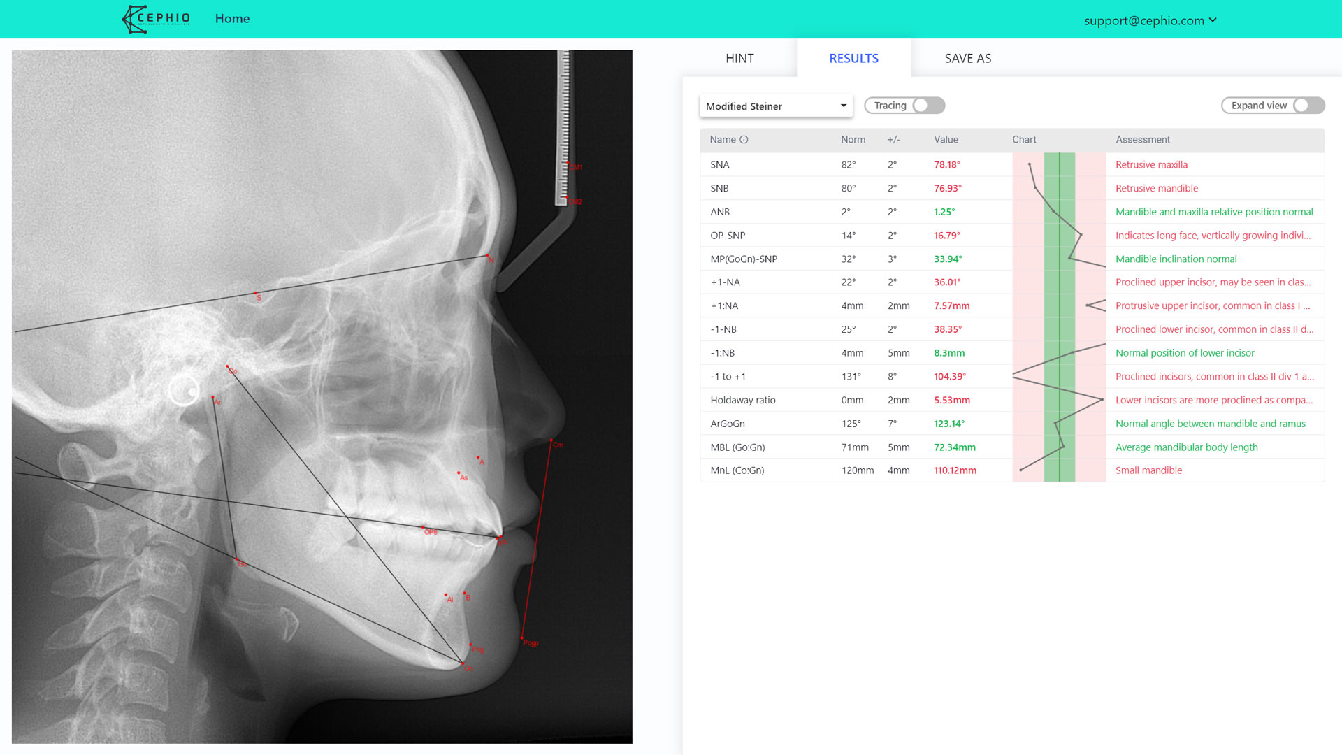
To do that, we've analyzed the digital X-ray in Cephio.

The Analysis used during the tests is a slightly modified Steiner with three extra parameters:
- angle between Articulare, Gonion and Gnathion,
- length from Condylion to Gnathion,
- mandibular length from Gonion to Gnathion.
The Gonial angle is chosen because it is on the left bottom of a skull, and we wanted to include in our test at least one angle from this region.
The two lengths are chosen because they are one of the longest measurements in cephalometry, and we've expected significant distortion effects in these parameters.
Pictures of X-rays
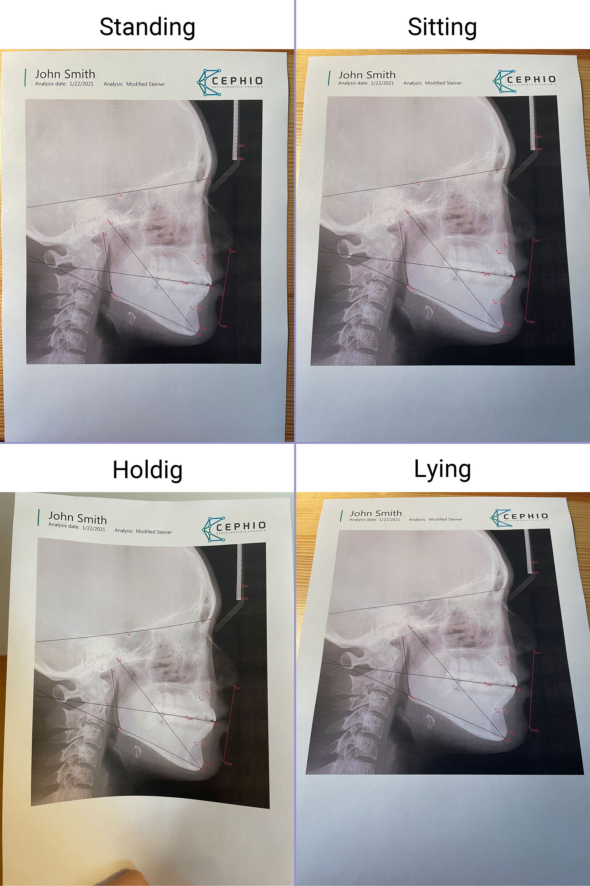
We've printed the reference analysis and made four pictures in the most common perspectives.
Note: We've used an iPhone 12 main camera. If you're using a different camera your results might slightly differ.
We've called them as follows:
- Standing - stand up and try to take a picture directly from the top,
- Sitting - sit straight in a chair and take a picture with a little perspective,
- Holding - hold analysis in hand with a small paper bending at the bottom,
- Lying - sit comfortably in a chair and take a picture with a significant perspective.

The idea of the test is quite simple.
We are going to investigate how the cephalometric analysis results depend on the perspective. Once we place the landmarks in the same locations as originally, we should get the same results unless there is a distortion.
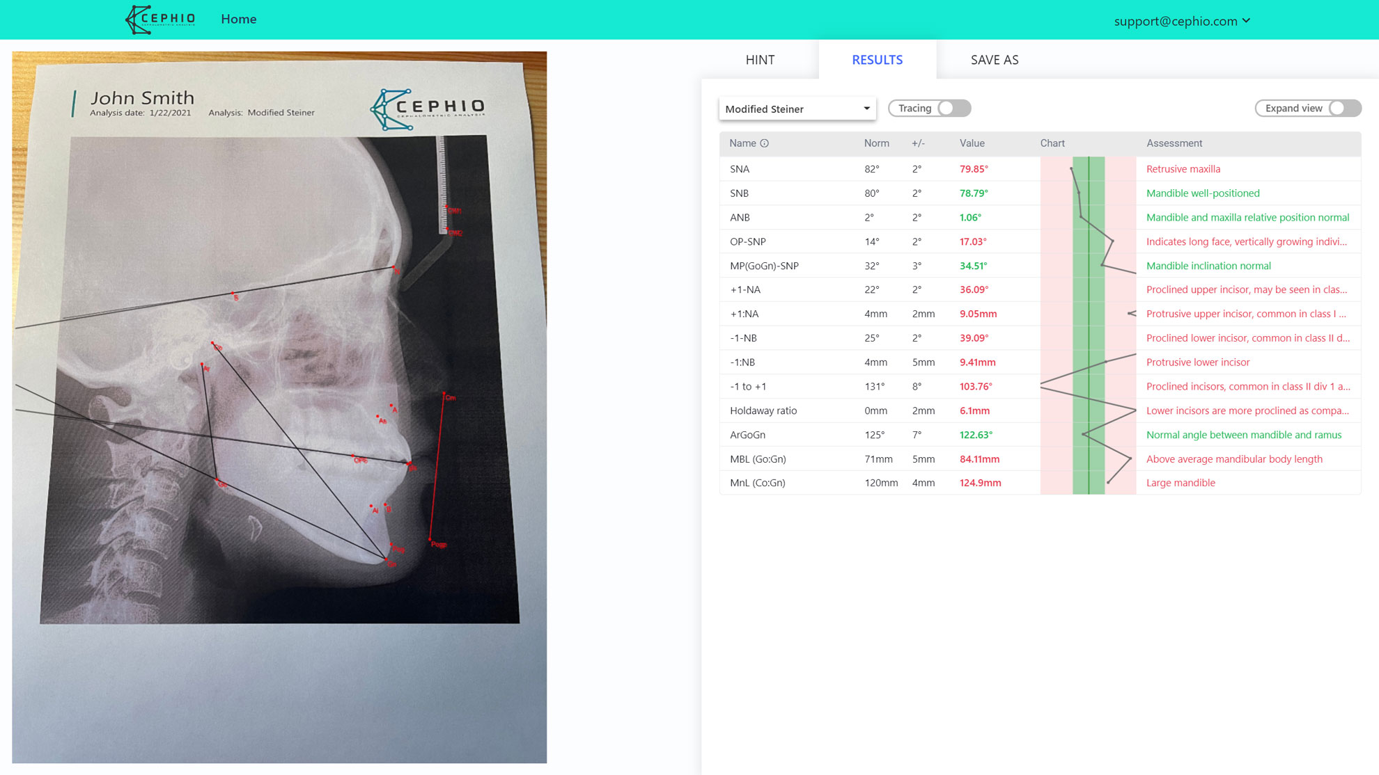
Here is an example of the "Sitting" picture. In the same way, we've done the remaining analyses.

Results
And now the most important part - How the results differ between pictures.
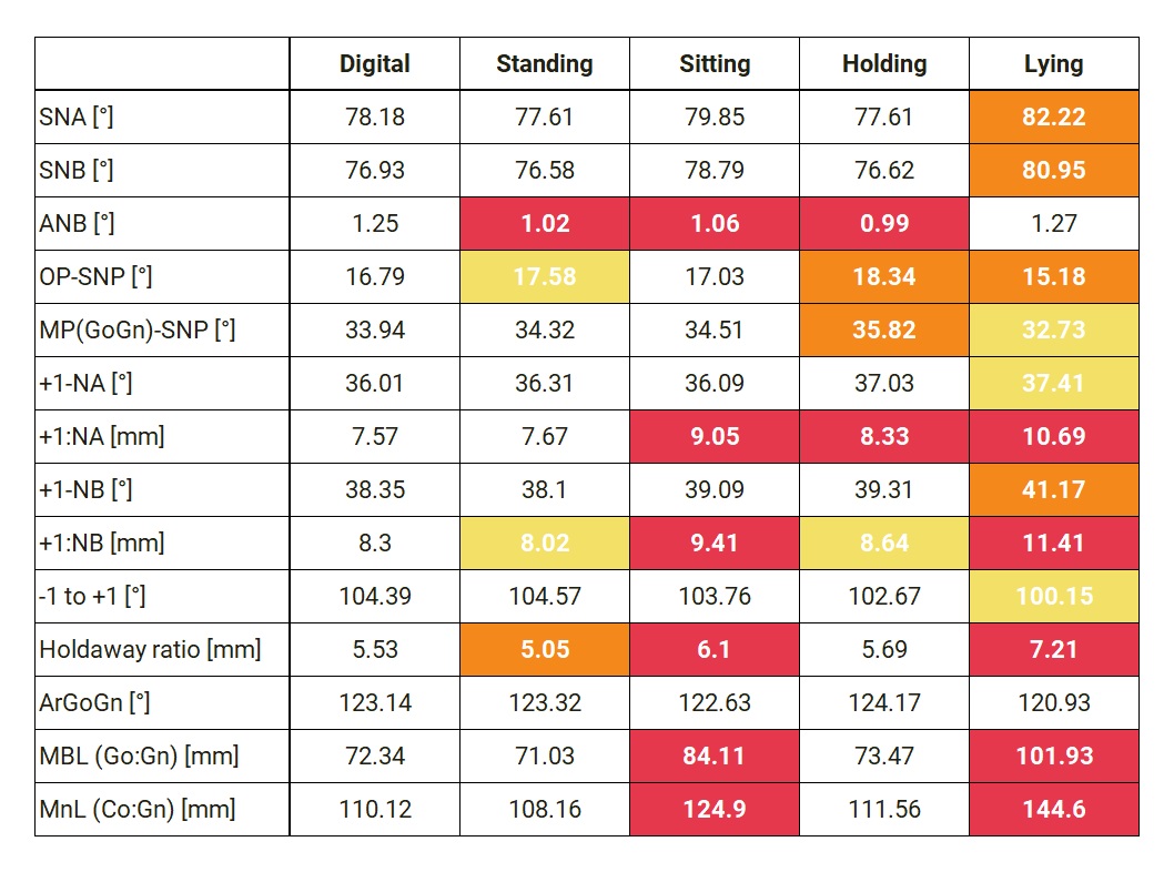
Deviations from the digital analysis were calculated and grouped into four classes:
- white: the difference is less than 3%,
- yellow: the difference is between 3-5%,
- orange: the difference is between 5-10%,
- red: the difference is more than 10%.

The worst results are for "Sitting" and "Lying" pictures. Unfortunately the "Sitting" perspective might be quite common.
The results which are the most distorted are the measurements. It is because, to calculate a distance in millimeters, we need to know two lengths:
- length in pixels of one centimeter,
- length in pixels of a measured distance.
Due to perspective, as the ruler is at the top, it is slightly "shrank", and the distances which are at the bottom are "stretched". These two distortions sum up together, resulting in a significant inaccuracy of over 1cm in mandibular length (Go:Gn)!!!
Surprisingly good results gave a "Holding" picture. This might indicate that the most significant distortions are caused by perspective - when the picture is not directly taken from the top.
The best results are as expected for the "Standing" picture, but keep in mind that they are still not 100% accurate.
It is also worth noting that differences in ANB angle are mainly because of human error. In this case, a 10% change (0.1 degrees) is caused by almost invisible displacement of landmarks.
Final thoughts
- The most fragile parameters are the measurements due to the propagation of errors (ruler + measured distance)
- Even a little perspective can distort an image leading to inaccurate results.
- Once you have to analyze a picture, try to capture it directly from the top or scan it.